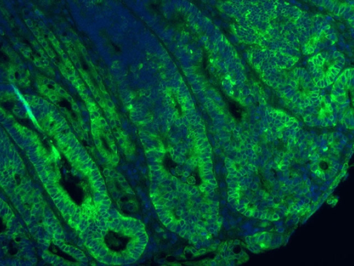
An intricate cellular dance during embryonic development is critical for normal bowel growth, a new Michigan Medicine study in mice finds.
7:00 AM
Author |

High-wire act: Motion of the nucleus of a dividing cell in the developing small intestine.
More than 40 percent of our small intestine develops before we are even born. In adulthood, the organ stretches more than three times the length of our bodies.
But problems with this process in utero can result in a rare but deadly condition known as congenital short bowel syndrome, which can lead to dehydration, malnutrition and weight loss during infancy.
LISTEN UP: Add the new Michigan Medicine News Break to your Alexa-enabled device, or subscribe to our daily audio updates on iTunes, Google Play and Stitcher.
A new study examining how the developing intestine grows in mice found a surprising sequence of cellular events akin to a death-defying, high-wire circus performance for the organ to reach a proper length. A lack of coordination could have dire consequences.
Sha Wang, Ph.D., and her Michigan Medicine colleagues were able to witness this intricate choreography in real-time in mouse gut cells.
The small intestine, or midgut, starts out as a smooth tube that undergoes rapid growth. Under a microscope, the cells in a thin layer lining the tube — known as epithelial cells — appear to be stacked in layers.
Previously, scientists believed that the tube grew longer via the movement of cells from these apparent stacks into one continuous layer.
"If you think about it, if the midgut lengthened through the convergence of cells, it would sacrifice girth (get thinner). But the tube gets wider," says Wang, a research fellow in the Department of Cell and Developmental Biology at the University of Michigan.
Instead, she and U-M cell biologist Deb Gumucio, Ph.D., and their team were able to show that this elongation is likely powered by rapid cell division. The results are published in Developmental Cell.
A high-stakes process
The team found that during the first phase of midgut development, cells are very tall and thin. The nucleus of each cell moves constantly within the epithelial layer, from the base (or basal) surface to the top (or apical) surface.
MORE FROM THE LAB: Subscribe to our weekly newsletter
In other words, sort of like a trapeze artist climbing up to their starting platform.
"The movement is tied to cell division," says Wang. "Once the nucleus is at the top, the cell will undergo mitosis and split into two new cells."
What happens next is critical. The two new daughter nuclei must make it back to the basal layer in order to re-enter the cell cycle.
For one daughter cell, this step is simple: Its nucleus simply follows a long, thin tether called a basal process back down.
The other cell has a tougher job to do; it must extend an armlike protrusion called a filopodium in the right direction before its nucleus can follow that path back down to the basal surface.
Divide and prosper
Upon closer inspection of the basal process, Wang noticed that this thin tether would often itself divide into two strands — one longer and tethered to the base and one short one that would retract and disappear.
Wang suspects the reabsorbed filament may play a role in dividing the cytoplasm, which makes up most of the body of each new daughter cell.
"The most interesting part is that the cells keep one basal process, which goes to one daughter cell," she says. "The other cell needs to find a connection back to the basal side. Even in the cases where both basal processes are kept, still only one daughter cell keeps them."
Wang hypothesizes that this is done purposefully to give one cell more flexibility for where it lands — but that cell also faces greater risk.
Telling up from down for the nontethered cell, as for a nontethered acrobat, is a matter of life and death.
How cells find their way
The team suspected that there was something serving as a directional cue to help newly formed cells know which way to extend their filopodium.
They settled on a protein known as WNT5A, which is known to help nerve cells grow similar projections called axons. This protein sends out a signal from the tissue, which the new untethered cell will extend toward to make its way back to the basal layer.
SEE ALSO: Study: Vaccine Suppresses Peanut Allergies in Mice
When Wang removed WNT5A, the cells were unable to point themselves in the right direction and died.
And that disconnect can have dire consequences.
Says Wang: "According to our model, even the death of just 10 percent of cells in the midgut can lead to shortened length."

Explore a variety of health care news & stories by visiting the Health Lab home page for more articles.

Department of Communication at Michigan Medicine
Want top health & research news weekly? Sign up for Health Lab’s newsletters today!
