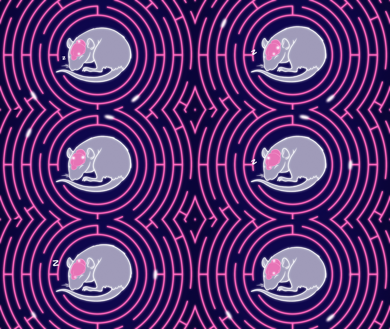
Sleep—or a lack thereof—has a dramatic effect on neurons in the hippocampus
11:00 AM
Author |

Imagine you’re a student, it’s finals week, and you’re preparing for a big exam: do you pull an all-nighter or do you get some rest?
As many a groggy-eyed person who’s stared blankly at a test knows, a lack of sleep can make it extraordinarily difficult to retain information.
Two new studies from University of Michigan uncover why this is and what is happening inside the brain during sleep and sleep deprivation to help or harm the formation of memories.
Specific neurons can be tuned to specific stimuli.
For example, rats in a maze will have neurons that light up once the animal reaches specific spots in the maze. These neurons, called place neurons, are also active in people and help people navigate their environment.
But what happens during sleep?
“If that neuron is responding during sleep, what can you infer from that?” said Kamran Diba, Ph.D., associate professor of Anesthesiology at U-M Medical School.
A study, summarized in the journal Nature and led by Diba and former graduate student Kourosh Maboudi, Ph.D., looks at neurons in the hippocampus, a seahorse shaped structure deep in the brain involved in memory formation, and discovered a way to visualize the tuning of neuronal patterns associated with a location while an animal was asleep.
A type of electrical activity called sharp-wave ripples emanate from the hippocampus every couple of seconds, over a period of many hours, during restful states and sleep.
Researchers have been intrigued by how synchronous the ripples are and how far they travel, seemingly to spread information from one part of the brain to another.
These firings are thought to allow neurons to form and update memories, including of place.
For the study, the team measured a rat’s brain activity during sleep, after the rat completed a new maze.
Using a type of statistical inference called Bayesian learning, they were for the first time able to track which neurons would respond to which places in the maze.
“Let’s say a neuron prefers a certain corner of the maze. We might see that neuron activate with others that show a similar preference during sleep. But sometimes neurons associated with other areas might co-activate with that cell. We then saw that when we put it back on the maze, the location preferences of neurons changed depending on which cells they fired with during sleep,” said Diba.
The method allows them to visualize the plasticity or representational drift of the neurons in real time.
It also gives more support to the long-standing theory that reactivation of neurons during sleep is part of why sleep is important for memories.
Given sleep’s importance, Diba’s team wanted to look at what happens in the brain in the context of sleep deprivation.
In the second study, also published in Nature, the team, led by Diba and former graduate student Bapun Giri, Ph.D., compared the amount of neuron reactivation—wherein the place neurons that fired during maze exploration spontaneously fire again at rest—and the sequence of their reactivation (quantified as replay), during sleep vs. during sleep loss.
They discovered that the firing patterns of neurons involved in reactivating and replaying the maze experience were higher in sleep compared to during sleep deprivation.
Sleep deprivation corresponded with a similar or higher rate of sharp-wave ripples, but lower amplitude waves and lower power ripples.
“In almost half the cases, however, reactivation of the maze experience during sharp-wave ripples was completely suppressed during sleep deprivation,” said Diba.
When sleep deprived rats were able to catch up on sleep, he added, while the reactivation rebounded slightly, it never matched that of rats who slept normally. Furthermore, replay was similarly impaired but was not recovered when lost sleep was regained.
Since reactivation and replay are important for memory, the findings demonstrate the detrimental effects of sleep deprivation on memory.
Diba’s team hopes to continue looking at the nature of memory processing during sleep and why they need to be reactivated and the effects of sleep pressure on memory.
Additional authors include Hiroyuki Miyawaki, Caleb Kemere, Nathaniel Kinshy, Utku Kaya and Ted Abel.
Citations:
“Retuning of hippocampal representations during sleep,” Nature. DOI: 10.1038/s41586-024-07397-x
“Sleep loss diminishes hippocampal reactivation and replay,” Nature. DOI: 10.1038/s41586-024-07538-2
Sign up for Health Lab newsletters today. Get medical tips from top experts and learn about new scientific discoveries every week by subscribing to Health Lab’s two newsletters, Health & Wellness and Research & Innovation.
Sign up for the Health Lab Podcast: Add us on Spotify, Apple Podcasts or wherever you get you listen to your favorite shows.

Explore a variety of health care news & stories by visiting the Health Lab home page for more articles.

Department of Communication at Michigan Medicine

Associate Professor
Want top health & research news weekly? Sign up for Health Lab’s newsletters today!


